| dc.contributor.advisor | Verchot-Lubicz, Jeanmarie | |
| dc.contributor.author | Mekuria, Tefera A. | |
| dc.date.accessioned | 2013-11-26T08:23:47Z | |
| dc.date.available | 2013-11-26T08:23:47Z | |
| dc.date.issued | 2007-07 | |
| dc.identifier.uri | https://hdl.handle.net/11244/6745 | |
| dc.description.abstract | Scope and Method of Study: | |
| dc.description.abstract | The green fluorescent protein (GFP) was fused to Potato virus X (PVX) TGBp1, TGBp2, or coat protein (CP) genes and inserted into the plasmids next to a companion cell specific CoYMV promoter. These plasmids were used to prepare transgenic tobacco plants expressing PVX proteins specifically in the phloem. Transgenic tobacco plants expressing a GFP:GFP fusion were also prepared as a control. GFP fluorescence was used to visualize phloem translocation and exit of the fusion proteins. Immunofluorescence and immunogold labeling was used to characterize the sub-cellular distribution of the fusion proteins within phloem. | |
| dc.description.abstract | Findings and Conclusions: | |
| dc.description.abstract | Fluorescence due to GFP:TGBp1, GFP:TGBp2, GFP:CP, and GFP:GFP was seen in transgenic tobacco leaf veins and failed to spread to nonvascular tissue. However, following PVX inoculation of GFP:TGBp1 and GFP:CP transgenic plants fluorescence spread into nonvascular tissues. Thus the destination of GFP:TGBp1 and GFP:CP, but not GFP:TGBp2 and GFP:GFP was altered by the presence of the virus indicating that the TGBp1 and CP directly interact with the virus to unload from the veins. Further studies explored phloem translocation of proteins within the stem and petioles. In petiole cross sections all fusion proteins were seen in phloem companion cells, sieve elements, and phloem parenchyma indicating that the proteins spread from the companion cells into surrounding phloem tissues. Only GFP:TGBp1 was seen in xylem parenchyma indicating that TGBp1 has a specific ability to move extensively throughout the vasculature. Nontransgenic scions were grafted to transgenic rootstocks and after 28 days fluorescence was seen in scions indicating that all fusions were phloem mobile. | |
| dc.description.abstract | Immunogold labeling and transmission electron microscopy revealed GFP:TGBp1 and GFP:CP accumulated in the chloroplasts, plastids, vacuoles and cytoplasm in companion cells, and associated with plastids and P-proteins in sieve elements. GFP:TGBp1 expressing plants showed increased numbers of starch bodies in xylem parenchyma, pericycles and endodermal cells and fewer tracheary elements in petiole cross sections suggesting that TGBp1 affects vascular development and carbohydrate partitioning. | |
| dc.format | application/pdf | |
| dc.language | en_US | |
| dc.rights | Copyright is held by the author who has granted the Oklahoma State University Library the non-exclusive right to share this material in its institutional repository. Contact Digital Library Services at lib-dls@okstate.edu or 405-744-9161 for the permission policy on the use, reproduction or distribution of this material. | |
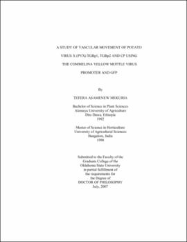
| dc.title | Study of vascular movement of Potato virus X (PVX) TGBp1, TGBp2 and CP using the Commelina yellow mottle virus promoter and GFP | |
| dc.contributor.committeeMember | Damicone, John P. | |
| dc.contributor.committeeMember | Melouk, Hassan A. | |
| dc.contributor.committeeMember | Anderson, Michael P. | |
| osu.filename | Mekuria_okstate_0664D_2355 | |
| osu.accesstype | Open Access | |
| dc.type.genre | Dissertation | |
| dc.type.material | Text | |
| thesis.degree.discipline | Entomology and Plant Pathology | |
| thesis.degree.grantor | Oklahoma State University | |
