| dc.contributor.author | Chakraborty, Songjukta | |
| dc.contributor.author | Muia, Joshua | |
| dc.contributor.author | Platt, Anna | |
| dc.contributor.author | Raju, Bhuvanesh Kumar | |
| dc.contributor.author | Miranda, Frida | |
| dc.contributor.author | Momani, Thomas | |
| dc.contributor.author | Ayomide, Ishola | |
| dc.date.accessioned | 2023-11-02T20:46:40Z | |
| dc.date.available | 2023-11-02T20:46:40Z | |
| dc.date.issued | 2023-02-17 | |
| dc.identifier | ouhd_Chakraborty_determinationofpresenceof_2023 | |
| dc.identifier.citation | Chakraborty, S., Muia, J., Platt, A., Raju, B. K., Miranda, F., Momani, T., and Ayomide, I. (2023, February 17). Determination of presence of ADAMTS7 in several human cell lines. Poster presented at Research Week, Oklahoma State University Center for Health Sciences, Tulsa, Ok. | |
| dc.identifier.uri | https://hdl.handle.net/11244/339913 | |
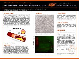
| dc.description.abstract | Background: According to studies, ADAMTS7 is associated with coronary atherosclerotic diseases. To understand the regulation of ADAMTS7, we have cultured the following laboratory available human cell lines to 3 or 4 passages: Hela, Human gingival fibroblast (HGF-1), Liver hepatocellular carcinoma (HepG2), and Human umbilical vein endothelial cells (HUVEC). These passage numbers are important because genetic and phenotypic changes will be less during early passages. After having 70 to 80% confluency, media was changed to Freestyle media and harvested after 48 hours of conditioning to do Western blotting. During the Western blot technique, primary antibody will bind with ADAMTS7 if this enzyme is present in the specific cell culture media. This process will help to understand the cell type and the amount of ADAMTS7 present in the cells. Method: We took the cell lines such as Hela, Human gingival fibroblast (HGF-1), Liver hepatocellular carcinoma (HepG2), Human umbilical vein endothelial cells (HUVEC) from cryogenic liquid and grew into T-25 flasks in DMEM or F-12K media with additives respectively. Aftersubculturing 3 to 4 passages into the T-150 flask, we replaced themedia with Freestyle media when the cells are 70 to 80% confluent. We harvested the media, added biotin to 10ml, and took samples of the biotinylated and non-biotinylated media for Western blotting. Then 25 ml conditioned media was concentrated by using viva spin columns and a sample of the concentrated media was taken for Western blotting. Result and Conclusion: Western blotting with the concentrated, unconcentrated and biotinylatedmedia to determine the expression of ADAMTS7 in specific human cell lines. Additionally, we will study that which cell lines are appropriate for ADAMTS7. We will further study the localization of ADAMTS7 by using imaging and microscopy. | |
| dc.format | application/pdf | |
| dc.language | en_US | |
| dc.publisher | Oklahoma State University Center for Health Sciences | |
| dc.rights | The author(s) retain the copyright or have the right to deposit the item giving the Oklahoma State University Library a limited, non-exclusive right to share this material in its institutional repository. Contact Digital Resources and Discovery Services at lib-dls@okstate.edu or 405-744-9161 for the permission policy on the use, reproduction or distribution of this material. | |
| dc.title | Determination of presence of ADAMTS7 in several human cell lines | |
| osu.filename | ouhd_Chakraborty_determinationofpresenceof_2023.pdf | |
| dc.type.genre | Presentation | |
| dc.type.material | Text | |
| dc.subject.keywords | atherosclerosis | |
| dc.subject.keywords | western blotting | |
| dc.subject.keywords | biotin | |
| dc.subject.keywords | human cell culture | |
