| dc.description.abstract | Introduction: Most schwannomas, other than those associated with neurofibromatosis, occur in the para-pharyngeal space often originating from Cranial nerves (CN) 9-12 or the sympathetic chain. Although cervical sympathetic chain (CSC) schwannomas are uncommon, they are known for their ability to mimic the physical and radiologic findings of carotid body tumors. Treatment is surgical resection. Post-operative complications involved with removing the CSC include Horner’s syndrome (almost inevitable) and First Bite Syndrome.
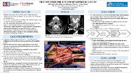
Case Description: An 80-year-old female presented with a 7-year history of an enlarging right neck mass followed serially over time with moderate growth. The patient complained of locally compressive symptoms including bilateral neck pain, dizziness, dysphagia and hoarseness. The mass was non-tender and firm with a 4-5 cm diameter oriented vertically. Flexible fiber-optic laryngoscopy revealed R vocal cord (VC) sluggishness with poor abduction. CT demonstrated the mass as measuring 3.6 x 3.5 x 4.9 cm with close adherence to the right carotid artery. Biopsy was non-diagnostic. The patient consented to mass excision and right neck dissection that was performed by an otolaryngologist and cardiovascular surgeon. The right-sided mass was free from the jugular vein, carotid artery, and CN 10. It measured 5.7 cm in the largest dimension and was attached to a nerve bundle, possibly the CSC. Histopathologic examination revealed the mass to be likely consistent with a schwannoma.
On post-operative day #1, the patient had normal tongue mobility, intact CN 7 function, slight ptosis of the right eye and symmetric pupils without miosis. Fiber-optic laryngoscopy revealed subtle right VC weakness that improved by post-operative day #5. Approximately one month later, the patient complained of discomfort in her mouth and jaw on initiating a meal. Following right parotid gland ultrasound examination, the patient was diagnosed with First Bite Syndrome. Botulinum toxin was injected within the parotid gland for symptom management.
Discussion: In First Bite Syndrome, pain in the parotid area can be severe with the first bite of food. With subsequent bites, the pain often decreases. It is currently thought this is likely due to sympathetic denervation of the parotid gland due to severing of the CSC resulting in hypersensitivity of the sympathetic receptors. A majority of these symptoms resolve over time. There is evidence that Botulinum toxin type A injection causes improvement of symptoms by inducing parasympathetic nerve paralysis of the parotid gland. This can also minimize salivation by reducing the hyper-stimulation and exaggerated myoepithelial cell contraction. | en_US |