| dc.description.abstract | INTRODUCTION: Fusobacterium nucleatum is an anaerobic, non-spore forming, gram-negative bacilli that is commonly found in soil, respiratory tracts of animals, and can be isolated from oropharyngeal specimens of healthy people. Fusobacterium spp. infections of dental plaque are most common in adolescents and young adults and may lead to periodontal disease. Fusobacterium spp. are capable of causing invasive disease commonly associated with otitis media, tonsillitis with Lemierre syndrome, gingivitis, and oropharyngeal trauma.
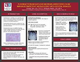
CASE DESCRIPTION: A previously healthy 16-year-old, Hispanic male initially presented with a three day history of fevers, chest pain, and lower back pain and acutely developed headache, neck pain, and vomiting. Broad spectrum empiric antibiotics were promptly started. Cerebral spinal fluid (CSF) was concerning for bacterial meningitis with white blood cell (WBC) count of 18,440 with 91% segmented neutrophils, red blood cells (RBC) 1260, Glucose 34, and Protein 222. Pre-treated CSF Gram stain and culture were negative. MRI of the brain showed numerous ring-enhancing lesions within bilateral cerebral hemispheres and brainstem concerning for multiple cerebral abscesses versus Neurocysticercosis (NCC). Fungal studies and immune studies including HIV and TB testing were all negative. Neuroimaging was reviewed with national NCC experts who did not feel the imaging was consistent with NCC. Serum serology sent to CDC for Neurocysticercosis testing ultimately returned negative. CSF 16s PCR analysis for fungal and broad range bacterial pathogens resulted as Fusobacterium nucleatum. Triple antibiotic therapy with ceftriaxone, vancomycin, and metronidazole was continued for eight weeks. Repeat CSF analysis showed remarkable improvement with WBC 169, RBC 146, Glucose 29, and Protein 68. Repeat MRI of the brain after finishing antibiotic therapy showed ring-enhancing lesions remarkably decreased in size and number with no new lesions demonstrated.
DISCUSSION: This case illustrates a very rare cause of brain abscesses in an immunocompetent adolescent with dental braces as the probable source of oropharyngeal infection. Further discussion with family and patient indicated that his oral hygiene routine was poor which likely lead to periodontal disease and ultimately dissemination of the organism to the brain. The diagnosis was ultimately established by 16s PCR analysis. The combination therapy with Metronidazole and Beta-lactams is recommended for an invasive disease. Because the patient was presumed to have meningitis, he received broad spectrum antibiotics which sterilized his CSF culture. This intriguing case of an uncommon cause of brain abscesses prompts further investigation of the role of Fusobacterium nucleatum causing disseminated disease in a healthy population. | en_US |

