| dc.description.abstract | INTRODUCTION: Histoplasma capsulatum is a thermally dimorphic fungi that is highly endemic in the central and eastern United States. The fungus lives in soil contaminated by bird or bat droppings and is aerosolized and inhaled causing infection. Histoplasmosis is generally a self-limited disease. Most symptomatic patients have acute pulmonary histoplasmosis while immunocompromised patients can have severe pulmonary or disseminated infection. This case emphasizes the importance of considering disseminated histoplasmosis in patients with a fever of unknown etiology.
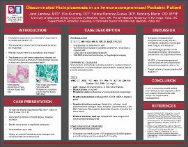
CASE DESCRIPTION: A 10-year-old female with Juvenile Dermatomyositis, managed with mycophenolate and methotrexate, presented to the Pediatric Emergency Center with ten days of fever (T max of 103.7°F) and symptoms of myalgia, fatigue, and anorexia. She had no recent travel or significant exposures and immunizations were up to date. Labs were remarkable for a WBC of 2.7 with absolute neutrophil count of 1100, hemoglobin of 11.9 and platelets of 79. Peripheral smear was consistent with mild leukopenia and marked thrombocytopenia. CRP was elevated to 2.46 and ALT to 69. Chest x-ray was negative for infectious process and blood cultures showed no growth. Due to persistent fevers, infectious work-up was broadened to include testing for common respiratory and gastrointestinal pathogens, Cytomegalovirus, Epstein-Barr virus, Aspergillus, Bartonella, Brucella, Francisella tularensis, Mycoplasma, and tick-borne illnesses, with all results negative. Due to worsening pancytopenia, a bone marrow biopsy was performed which revealed non-caseating granulomas with fungal yeast forms consistent with Histoplasma capsulatum. Histoplasmosis urine and serum antigen tests were positive, as was bone marrow culture. She completed a two-week course of intravenous amphotericin B while inpatient, with continued oral itraconazole for 12 months.
DISCUSSION: Disseminated histoplasmosis is acquired by inhaling fungal spores in endemic areas and presents with symptoms of prolonged fever, fatigue, anorexia and hepatosplenomegaly. Common laboratory findings include pancytopenia, transaminitis and hyperbilirubinemia. Typical radiologic findings include diffuse reticulonodular, interstitial, or military infiltrates, however 40–50% of immunocompromised pediatric patients with disseminated disease have negative chest x-rays. Tissue culture demonstrating typical fungal yeast forms is definitive for diagnosing histoplasmosis. Treatment with intravenous amphotericin B is given for a minimum of 2 weeks, contingent on clinical response. When clinical improvement is demonstrated, oral itraconazole is given for 12 months.
Timely diagnosis of disseminated histoplasmosis can be challenging due to its heterogenous clinical presentation. This case illustrates disseminated histoplasmosis as an important differential diagnostic consideration especially in immunocompromised patients presenting with systemic illness. | en_US |