| dc.contributor.advisor | Grady, Brian P. | |
| dc.creator | Mondalek, Fadee George | |
| dc.date.accessioned | 2019-04-27T21:32:13Z | |
| dc.date.available | 2019-04-27T21:32:13Z | |
| dc.date.issued | 2009 | |
| dc.identifier | 99276548602042 | |
| dc.identifier.uri | https://hdl.handle.net/11244/318944 | |
| dc.description.abstract | Small intestinal submucosa (SIS) derived from porcine small intestine has been intensively studied for its capacity in repairing and regenerating wounded and dysfunctional tissues. However, SIS suffers from a large spectrum of heterogeneity in microarchitecture leading to inconsistent results. The goal of this study is to modify an already existing SIS biomaterial with poly(lactide-co-glycolide) nanoparticles (PLGA NPs) for better tissue regeneration. | |
| dc.description.abstract | Specifically, nanoparticles made from PLGA were introduced into the SIS with the intention of decreasing the heterogeneity and improving the regenerative capability of this biomaterial. As determined by scanning electron microscopy and urea permeability, the optimum NP size was estimated to be between 200-500 nm. The PLGA NPs embedded in the SIS reduced the permeability of SIS to urea by almost 2-fold and did not change the tensile properties of the modified SIS as compared to the native SIS. More importantly, PLGA NP modified SIS did not affect human microvascular endothelial cells (HMEC-1) morphology or adhesion, but actually enhanced their growth. | |
| dc.description.abstract | These NPs were also used to deliver hyaluronic acid (HA) to enhance the angiogenic potential of SIS. Hyaluronic acid is an essential part of the extracellular matrix (ECM). HA in biomaterials has been shown to help improve tissue regeneration and enhance angiogenesis. To target optimal delivery of exogenous HA, an understanding of the expression of HA and its receptors in regenerated rat bladder was investigated. Our studies indicate that expression of HA and HA receptors, CD44 and LYVE-1, were not detected in rat bladder augmented with SIS until day 28 post-augmentation and thereafter. | |
| dc.description.abstract | HA-PLGA NPs were formulated to reduce the porous structure of SIS. Elevated cell proliferation was observed in HMEC-1 cells treated with HA-PLGA NPs as well as in HA-PLGA modified SIS (HA-PLGA-SIS). The HA-PLGA-SIS exhibited significantly enhanced neo-vascularization in an in ovo chorioallantoic membrane (CAM) angiogenesis model. The regenerative capability of the newly fabricated HA-PLGA-SIS was tested in a large animal bladder augmentation model. Bladders regenerated with HA-PLGA-SIS had significantly higher vascular ingrowth as compared to unmodified SIS. Based on the consensus that angiogenesis is essential for tissue regeneration, HA-PLGA-SIS represents a new approach for modifying naturally derived biomaterials in regenerative medicine. | |
| dc.format.extent | 110 pages | |
| dc.format.medium | application.pdf | |
| dc.language | en_US | |
| dc.relation.requires | Adobe Acrobat Reader | |
| dc.subject | Biomedical materials | |
| dc.subject | Tissue engineering | |
| dc.subject | Nanoparticles | |
| dc.subject | Animal cell biotechnology | |
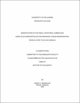
| dc.title | MODIFICATION OF THE SMALL INTESTINAL SUBMUCOSA USING PLGA NANOPARTICLES FOR ENHANCED TISSUE REGENERATION: FROM IN VITRO TO IN VIVO MODELS | |
| dc.type | text | |
| dc.type | document | |
| dc.thesis.degree | Ph.D. | |
| ou.group | College of Engineering::School of Chemical, Biological and Materials Engineering | |
